HYPOSPADIAS BEFORE AND AFTER PHOTOS
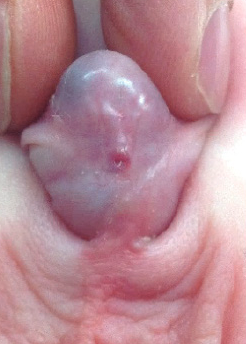
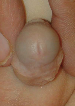
The picture to the left shows an infant patient with uncorrected distal hypospadias, while the picture to the right shows the appearance 6 months after his distal TIP repair with a circumcision.
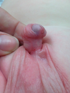
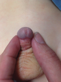
This infant was diagnosed with distal hypospadias as well as penile torsion. The picture to the left is before surgery while the picture to the right is 6 months after his distal TIP repair with circumcision.
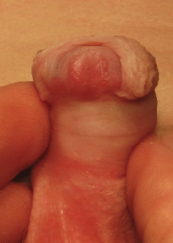
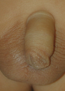
This infant has been diagnosed with distal hypospadias which can be seen in the left picture. The picture to the right of that is 8 months after a TIP repair with foreskin reconstruction (no circumcision).
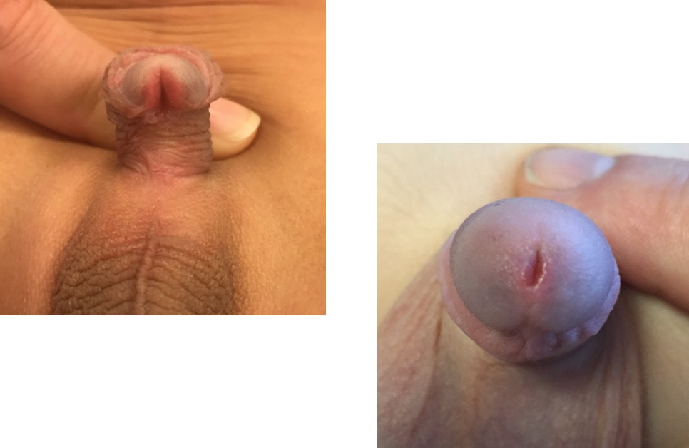
This patient had distal hypospadias, he received a single repair and the family desired a circumcision.
This patient was diagnosed with proximal perineal hypospadias with significant penis curvature that has been corrected with a staged repair STAG repair as an infant. This picture to the right is 3 months after the 2nd stage surgery.
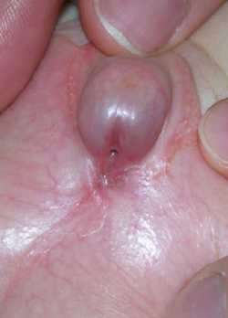
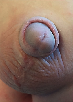
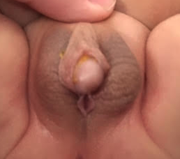
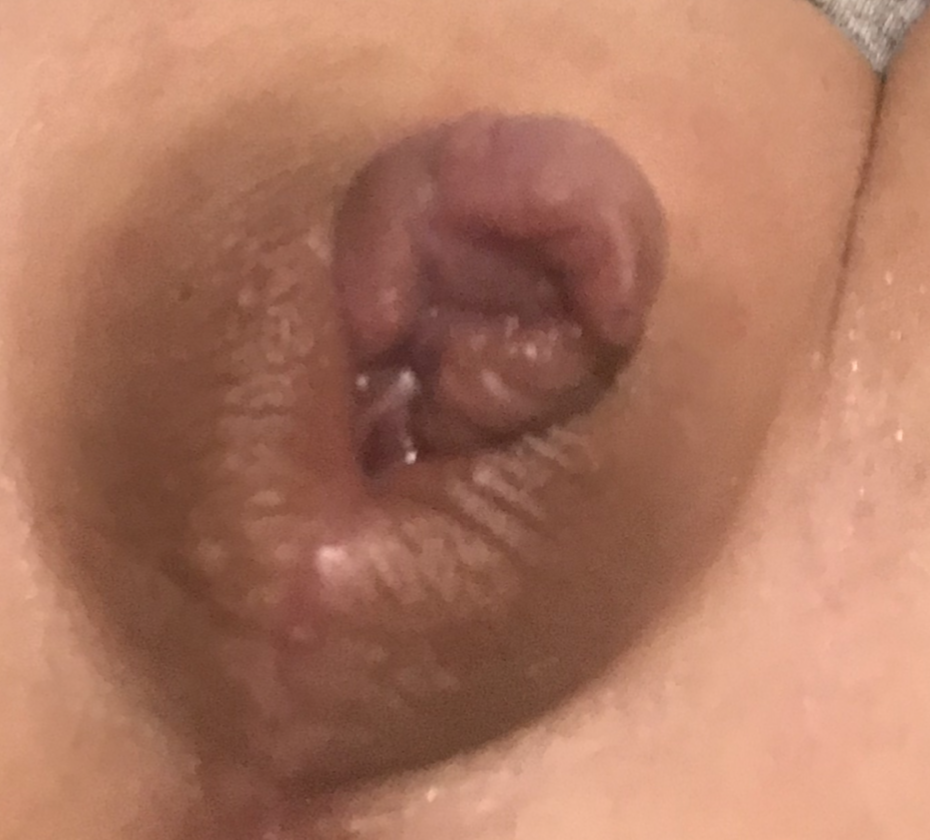
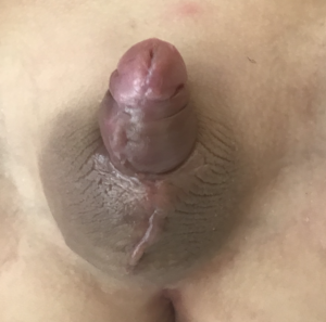
This patient presented to our office with proximal penoscrotal hypospadias and was repaired using a staged repair STAG repair with genital skin graft. The first picture is pre-operatively, the second picture is after the 1st stage surgery when the graft was placed, and the last picture is 3 weeks after the 2nd surgery.
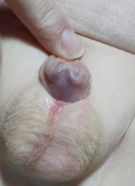
This infant was diagnosed with proximal hypospadias repairing a staged repair. The photo on the right was taken 2 weeks after his 2nd surgical repair.
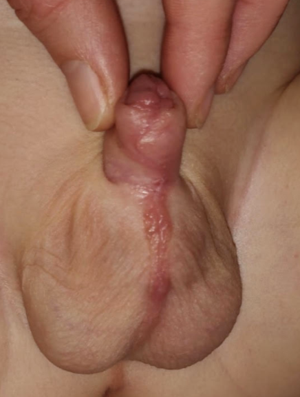
This patient presented to our office with proximal penoscrotal hypospadias requiring a staged repair STAG approach in order to correct. The picture in the middle was taken 6 months old and the picture to the right was taken 3 months after his 2nd surgery.
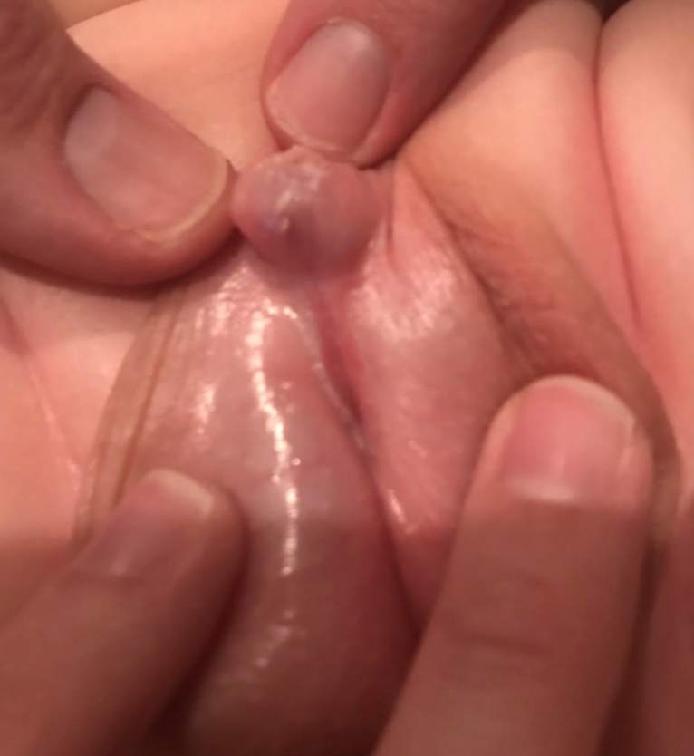
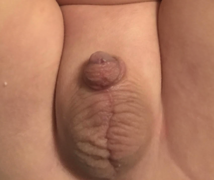
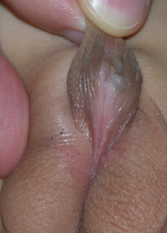
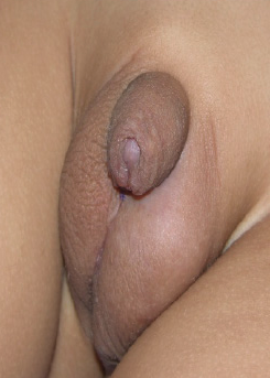
The picture to the left shows this kiddo who was diagnosed with proximal scrotal hypospadias as well as penis curvature. He was corrected using a staged repair STAG repair with oral graft due to the parents request for a more natural appearance (desired foreskin reconstruction - no circumcision).
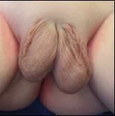
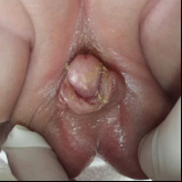
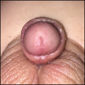
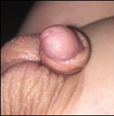
This patient was diagnosed with proximal perineal hypospadias (the first two pictures) and presented with significant penis curvature and penoscrotal transposition. This is an example of one of the most severe forms of hypospadias. The patient underwent a staged repair (two surgeries) STAG repair using a graft to correct thetransposition using incisions that are hidden within the natural skin creases. This technique gives a normal post-operative appearance and function. The last two pictures are 6 weeks after his 2nd surgery.
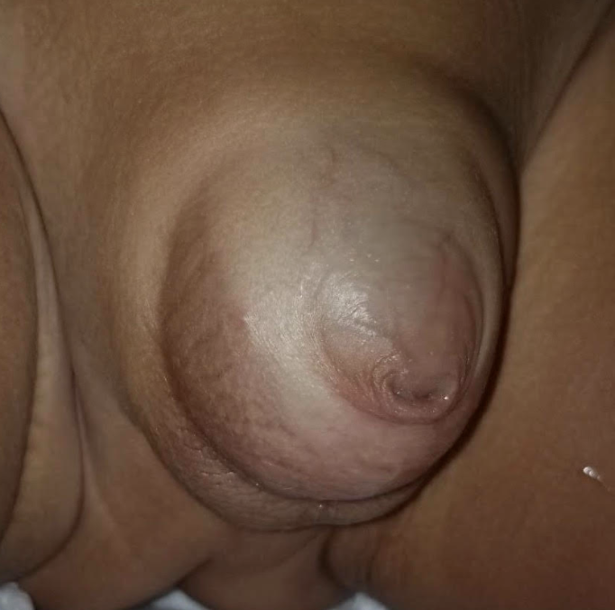
This infant came to us with a diagnosis of hypospadias, megaprepuce, and congenital concealed penis that required a staged repair repair for correction. The picture on the left was prior to surgery while the picture on the right was 4 weeks after the 2nd surgery.
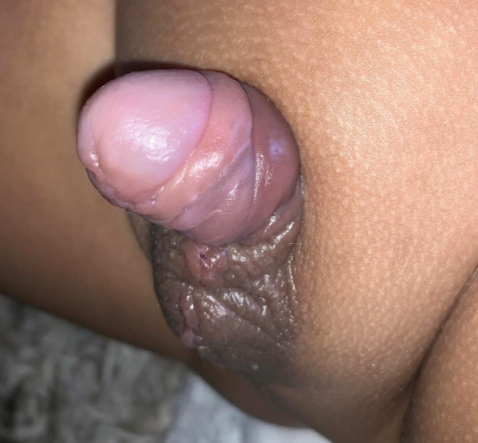

This child came to our office with chordee and scrotal hypospadias. He required a staged repair STAC repair.
The picture to the left shows this patients proximal penoscrotal hypospadias with significant penis curvature. His hypospadias was corrected with a staged repair grafting repair, and the picture to the right is 6 weeks after his 2nd surgery.
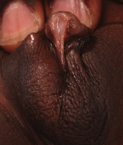
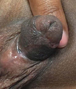
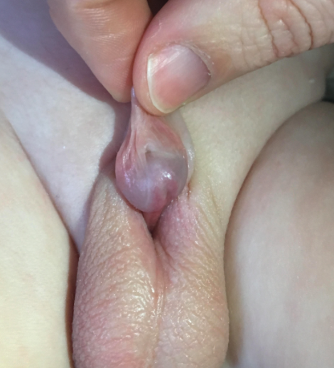
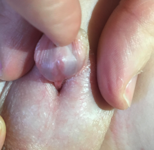
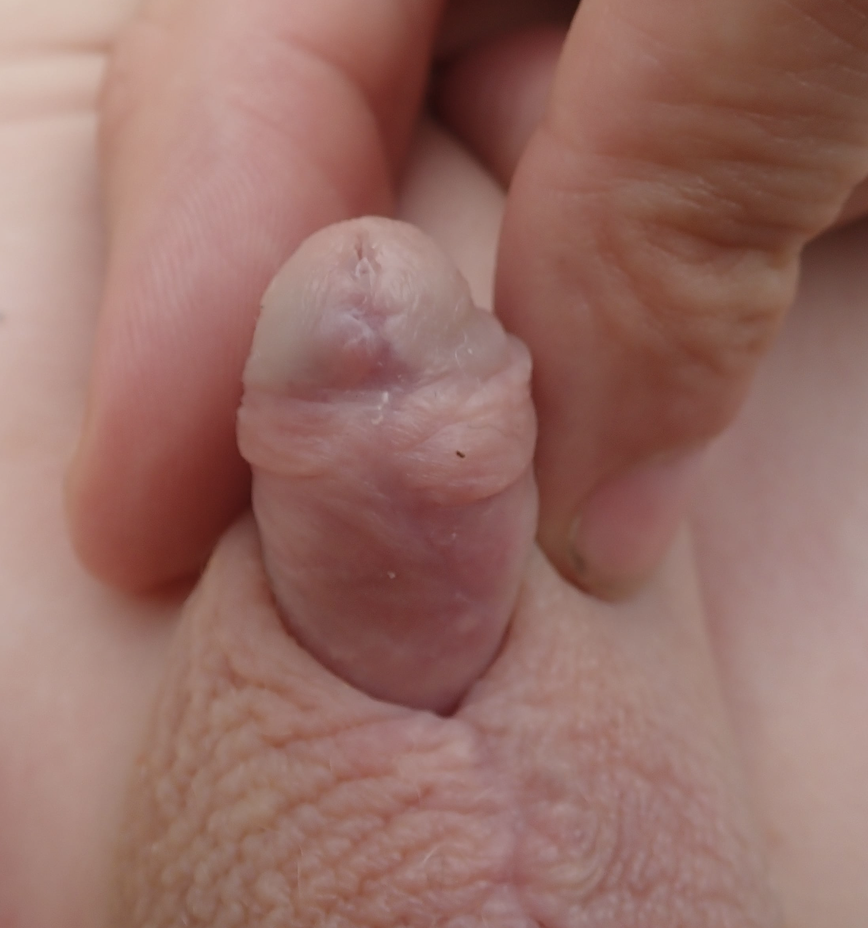
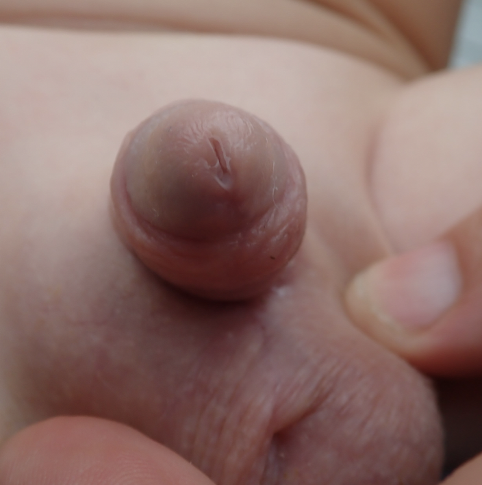
This infant patient presented to our office with proximal hypospadias requiring a staged repair STAC then STAG (straightening, lengthening, and lastly tubularizing) surgical approach to correct his hypospadias. The first two pictures show his pre-operative appearance while the next two pictures are 6 weeks after the staged repair surgical approach using oral graft.
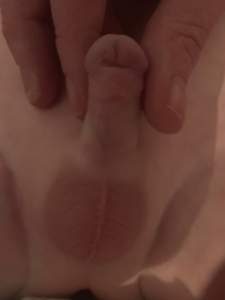
This patient had a failed distal TIP repair followed by glans dehiscence performed by another surgeon prior to coming to our office. In order to correct this a staged repair was performed. The picture to the right was taken 3 months after his 2nd surgical repair.
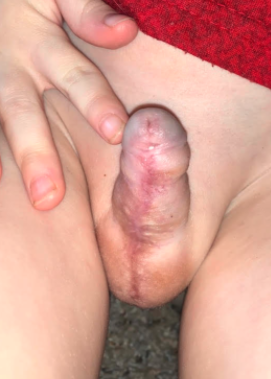
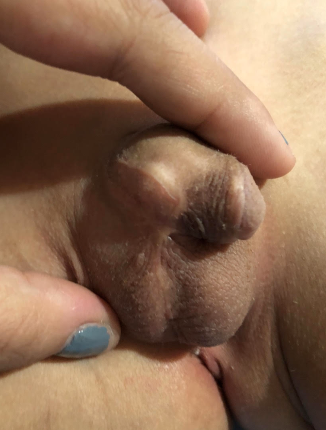
This patient came to our office after having two previously failed hypospadias repairs with another surgeon. It was determined that his proximal hypospadias repair needed to be corrected using a staged repair STAG repair approach. This picture to the right was taken 6 weeks after his 2nd surgical repair.
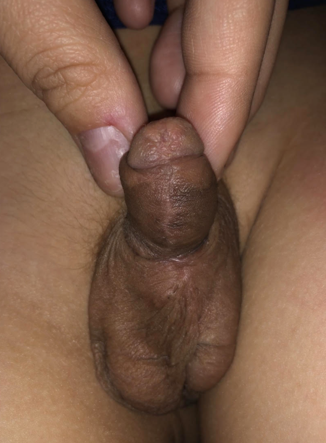
This patient had a Byars flap repair surgery with dorsal plication performed by another surgeon that healed with dehiscence of the wound. In order to correct his hypospadias a staged repair STAC with STAG repair needed to be performed. The initial pictures is before the staged repair approach while the second picture was taken 6 weeks after his 3rd surgery.
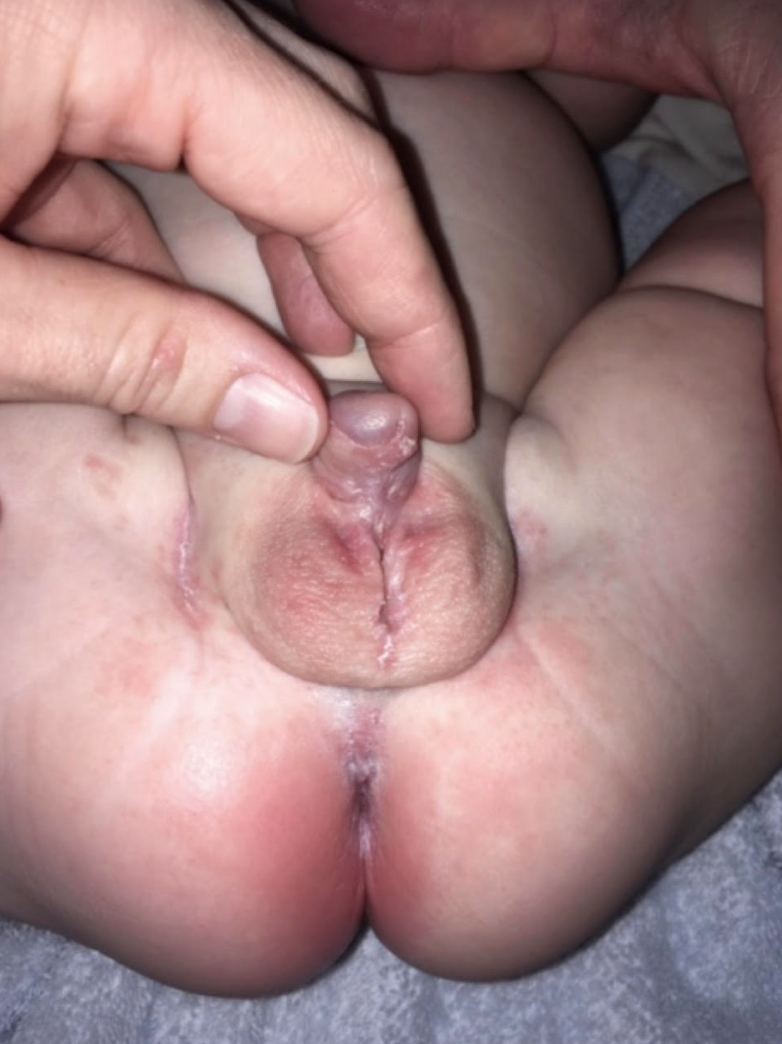
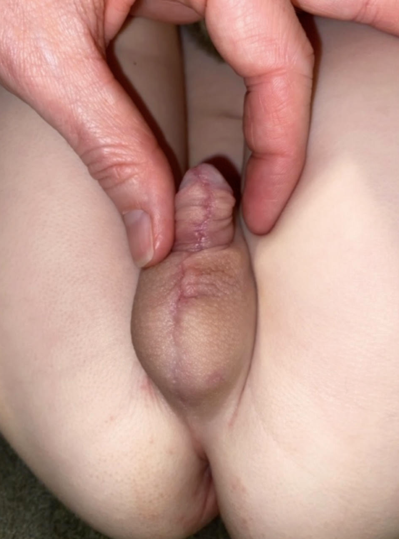
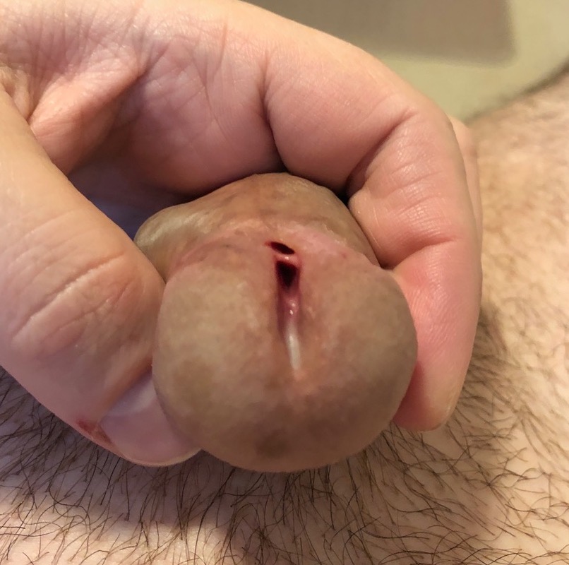
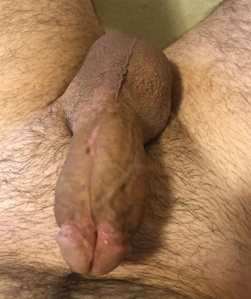
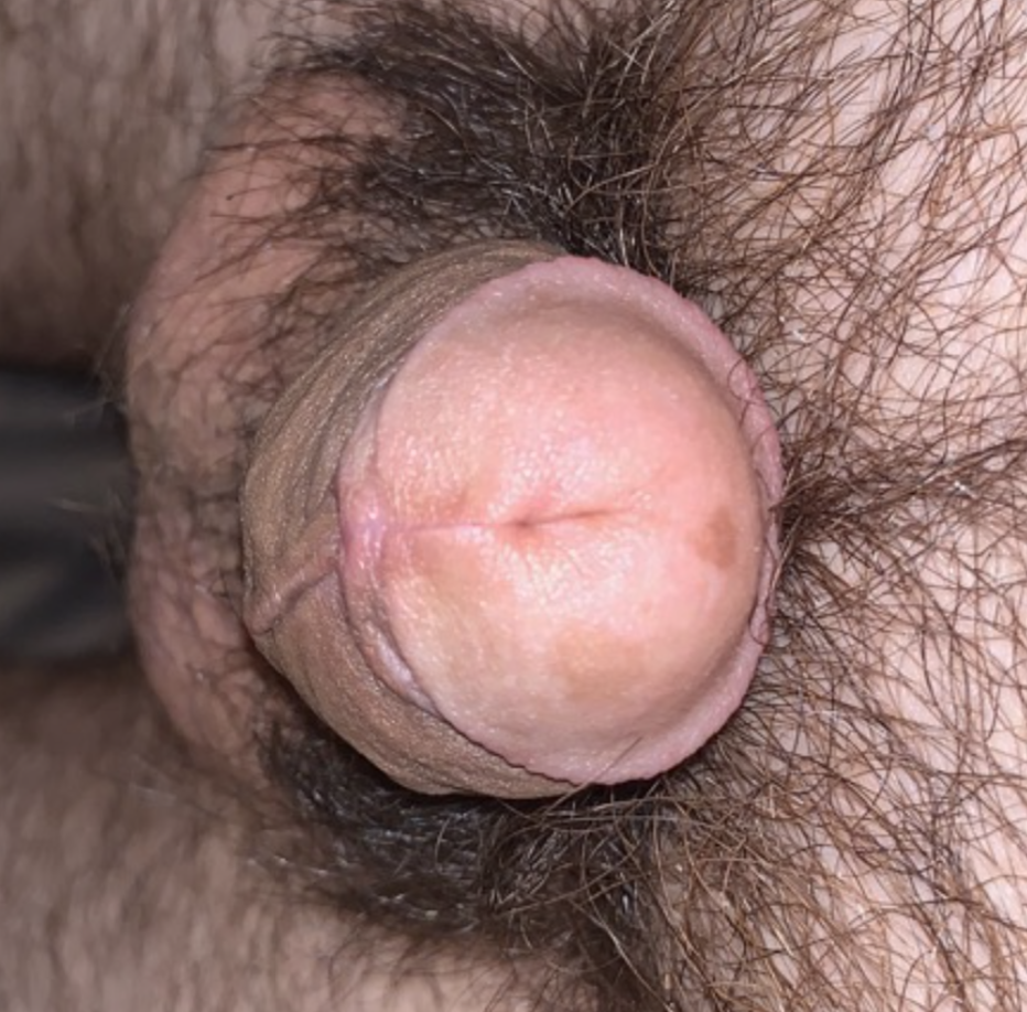
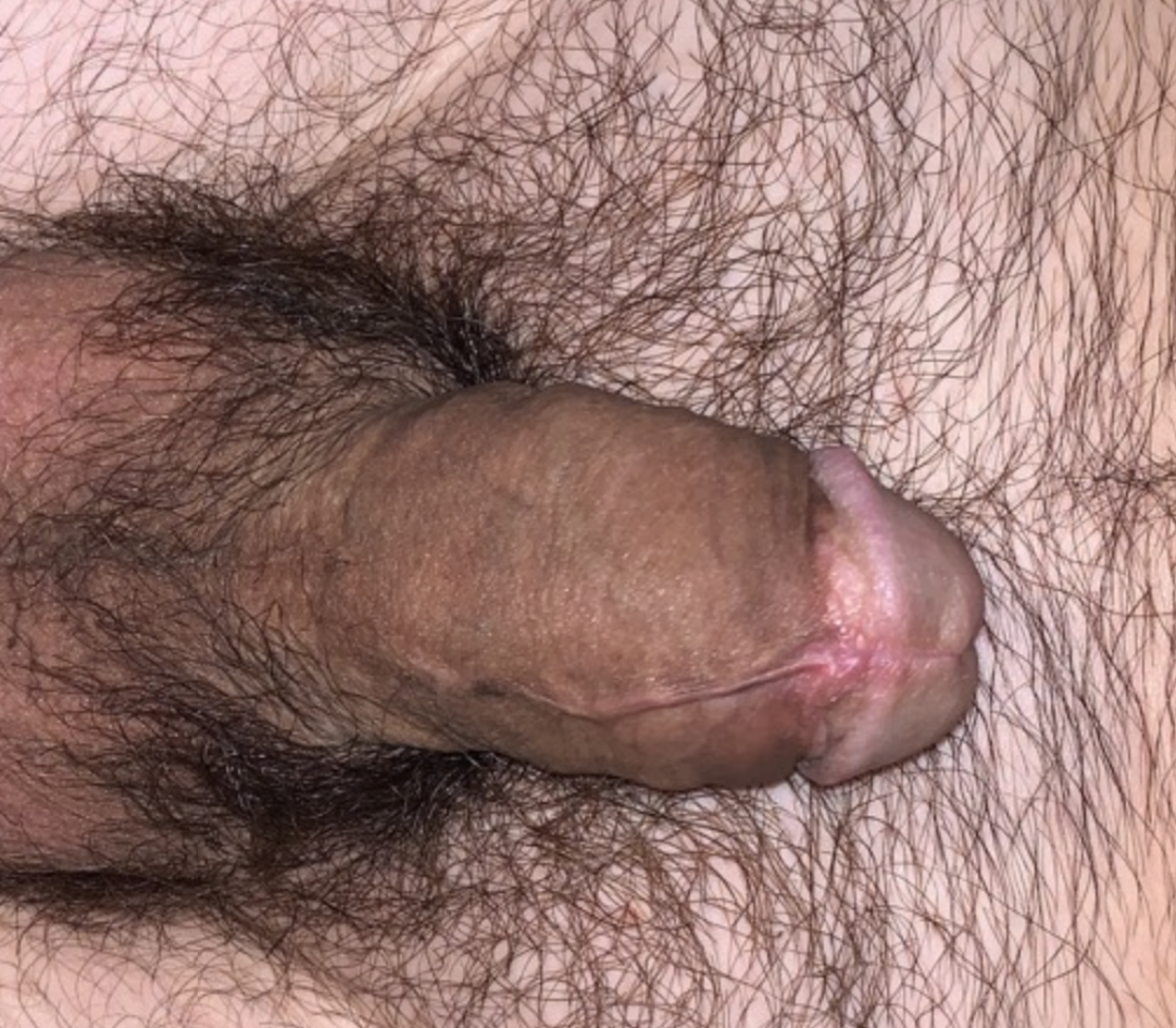
This adult patient suffered from distal hypospadias and spraying with urination. He was corrected using a 1-stage TIP repair followed with post-operative hyperbaric treatments. The two pictures to the right were taken 6 weeks after the TIP repair.
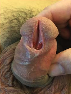
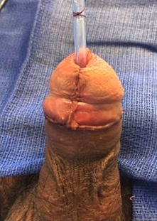
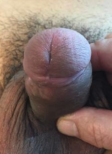
This adult patient was diagnosed with distal hypospadias and suffered from urine spraying. The first picture is pre-operatively, the second picture was immediately after surgery, while the last picture is 6 weeks after surgery.
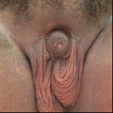
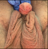
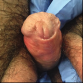
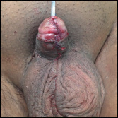
This Teen presented with a prior hypospadias repair using a flap-based approach by another surgeon. He has proximal hypospadias with penis and scrotal skin scarring which entrapped the penis, along with glans dehiscence and urinary spraying. Surgery was performed to remove the scar tissue,, unbury the penis, and correct eh glans dehiscence.
This teenage patient was referred to our office due to his glans dehiscence along with skin scarring after a proximal hypospadias repair by another surgeon. He was able to be repaired in a 1-stage TIP procedure with skin revision.
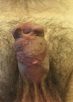
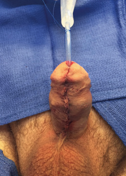
This adult patient presented with scarring of his glans as well as recurrent fistulas after a distal hypospadias repair from childhood by another surgeon. Our office corrected his distal hypospadias repair with glans contouring and hyperbaric oxygen treatments to promote skin healing.
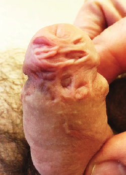
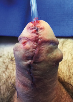
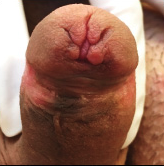
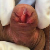
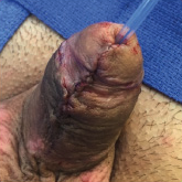
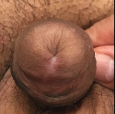
This patient underwent a ailed distal hypospadias repair that left deep cuts in the head of the penis which caused spraying of the urine stream in this young adult patient. The first two pictures show the appearance before surgery, the third is intraoperatively, and the last picture is post operatively. The last pictures shows the glans contouring with a near-normal appearance with marked improvement in urine spraying.
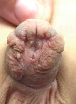
This patient presented with extensive glans scarring and complete wound dehisence after having a prior distal hypospadias repair by another surgeon. In order to correct this a TIP urethroplasty was performed along with glans sculpting and skin revisioning. The picture to the right is immediately after surgery.
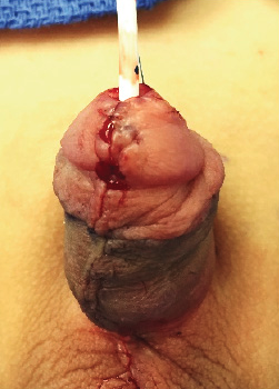
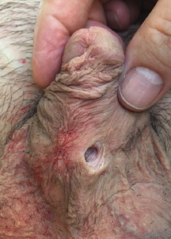
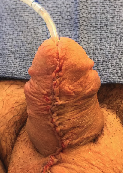
This adult patient presented to our office with a large fistula and unsightly skin following a proximal hypospadias repair performed at childhood by another surgeon. The picture to the left is before surgery while the picture to the right of that is immediately following the fistula corrective surgery with skin revision.
